
Research
PICSL develops computational methods for biomedical image analysis, including segmentation, registration, morphometry and shape analysis, and applies them across organ systems and imaging modalities.
Projects
2026
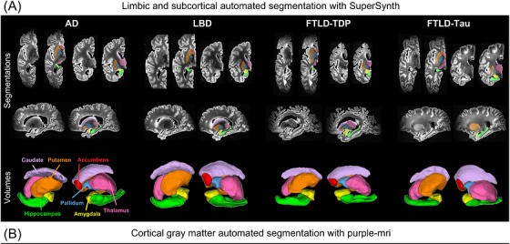
Postmortem MRI and neuropathology from 132 brain donors reveal disease- and region-specific links between subcortical and limbic atrophy and tau, α-synuclein and TDP-43. Published in Alzheimer’s & Dementia.
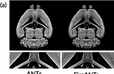
A training-free, GPU-accelerated registration algorithm from the Gee lab that is two orders of magnitude faster than ANTs on a GPU and robust across modalities, species and organs. Published in Nature Communications.
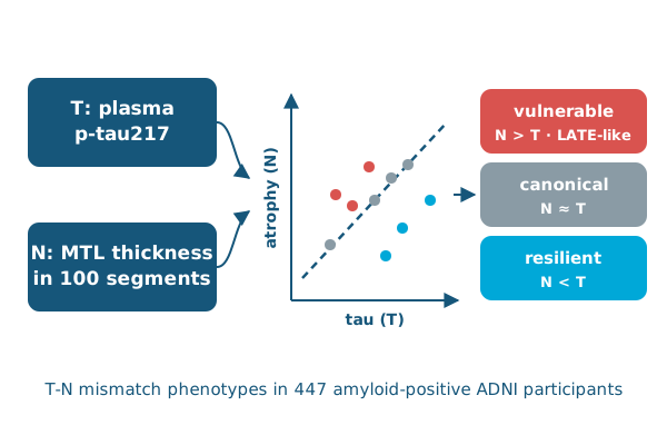
Mismatch between plasma p-tau217 and medial temporal lobe atrophy identifies vulnerable, canonical and resilient groups, including patients with likely LATE co-pathology. Published in Brain.
2025
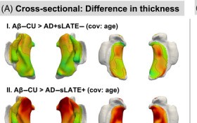
Lower-quartile hippocampal volume identifies Alzheimer’s patients with suspected LATE, who show more anterior MTL atrophy and faster decline. Published in Alzheimer’s & Dementia.
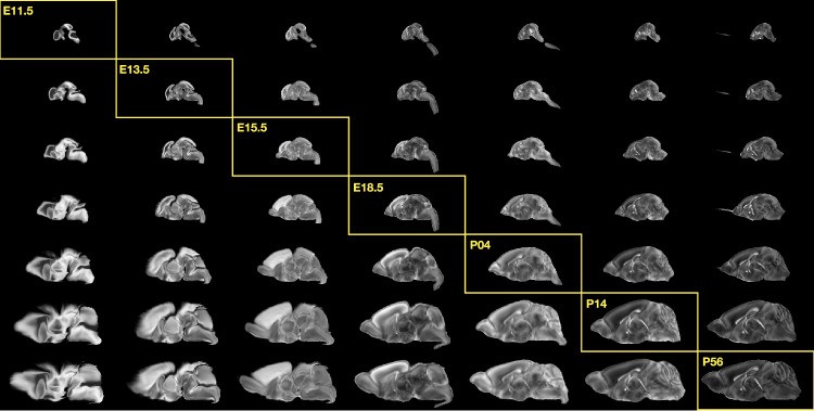
Open-source ANTsX pipelines align spatial transcriptomics, fMOST morphology, MRI and light-sheet data to common mouse brain atlases for the BRAIN Initiative. Published in Nature Communications.
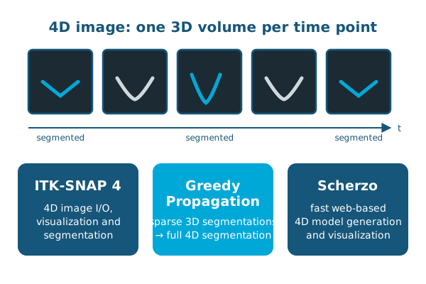
ITK-SNAP 4, Greedy Propagation and Scherzo bring native 4D segmentation, registration and visualization to cardiac imaging. Presented at FIMH 2025.
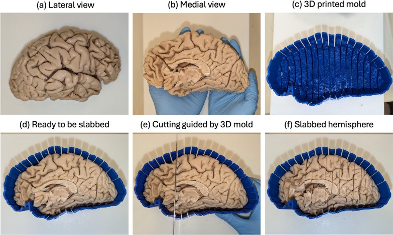
Patient-specific 3D-printed molds, MRI-to-histology registration and deep learning pathology scoring make large postmortem pathology-MRI studies practical. Published in Acta Neuropathologica Communications.
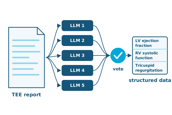
Consensus voting across five open-source LLMs extracts key measurements from intraoperative echocardiography reports with up to 99% accuracy. Published in the British Journal of Anaesthesia.
2024
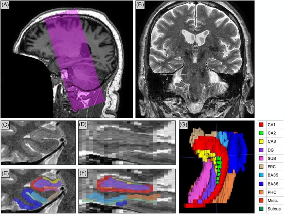
For ADNI’s 20th anniversary: why a dedicated hippocampal subfield MRI scan joined ADNI3, how it is analyzed, and what surface-based analysis reveals about tau-driven atrophy. Published in Alzheimer’s & Dementia.
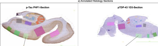
Deep learning quantification of p-tau and TDP-43 on histology, compared with antemortem MRI in 140 cases, outperforms standard pathology ratings at explaining atrophy. Published in Acta Neuropathologica.
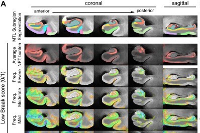
Ex vivo MRI and serial histology from 25 specimens map where tau tangles accumulate in the medial temporal lobe in 3D, informing early Alzheimer’s biomarkers. Published in Nature Communications.
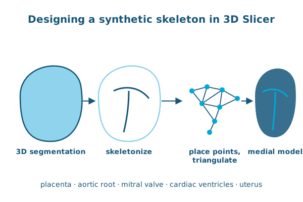
A 3D Slicer interface for designing synthetic skeletons makes deformable medial modeling practical for new anatomies, from the placenta to heart valves. Published in the Journal of Medical Imaging.
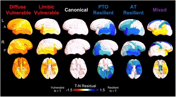
Regional mismatch between tau PET and atrophy groups Alzheimer’s patients by non-AD factors and predicts diverging outcomes; limbic mismatch tracks TDP-43 at autopsy. Published in Alzheimer’s & Dementia.
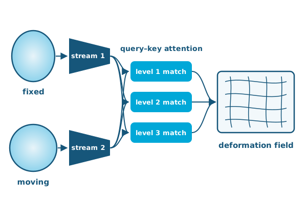
A dual-stream transformer network that explicitly matches features between images at multiple levels, achieving state-of-the-art unsupervised brain MRI registration. Published in IEEE Transactions on Medical Imaging.
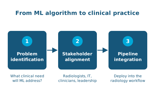
A step-by-step framework for implementing AI in clinical radiology: problem identification, stakeholder alignment and pipeline integration. Published in Radiology.
2023
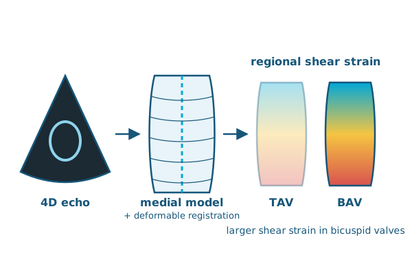
Medial modeling and deformable registration estimate aortic root strains in vivo, revealing larger shear strains in patients with bicuspid aortic valves. Published in Medical Image Analysis.
2022
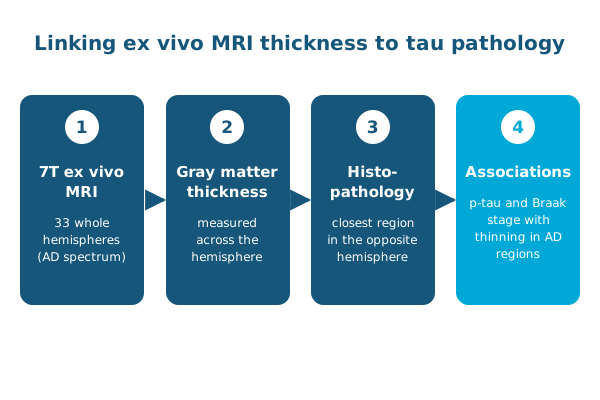
Pairing thickness from 7 tesla ex vivo MRI of 33 hemispheres with histopathology links tau pathology to tissue loss in key Alzheimer’s regions. Published in Alzheimer’s & Dementia.
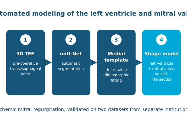
Deep learning segmentation and diffeomorphic medial modeling produce shape models of the left ventricle and mitral valve from pre-operative 3D echo. Published in Medical Image Analysis.
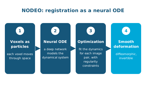
Treating voxels as particles in a dynamical system modeled by a neural ODE gives accurate diffeomorphic registration with built-in regularity constraints. Presented at CVPR 2022.
2021
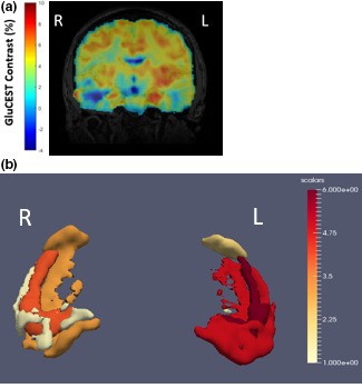
Volumetric GluCEST imaging at 7 tesla showed higher glutamate in the hippocampus on the side of seizure onset in MRI-negative temporal lobe epilepsy. Published in Brain and Behavior.
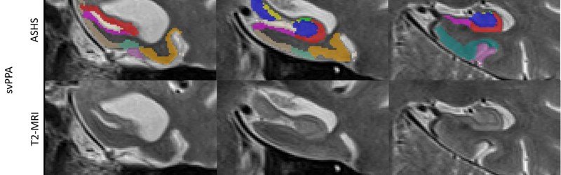
High-resolution T2 MRI and ASHS reveal perirhinal and CA1 atrophy in semantic variant primary progressive aphasia, with faster BA36 atrophy over time. Published in Neurobiology of Aging.
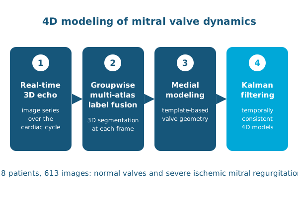
Multi-atlas segmentation, medial modeling and Kalman filtering produce temporally consistent 4D models of the mitral valve from real-time 3D echo. Published in Experimental Mechanics.
2020
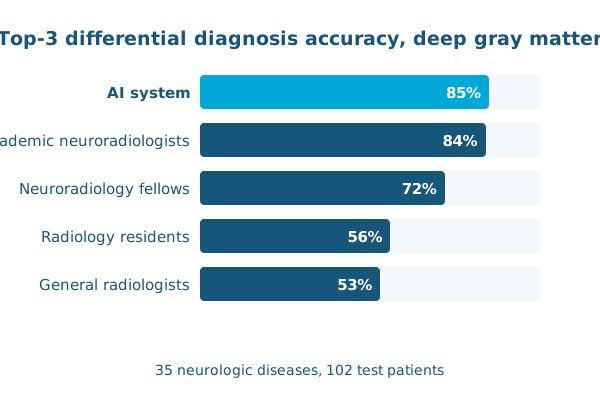
Deep learning feature extraction plus expert-derived Bayesian networks diagnosed 35 diseases of the deep gray matter at the level of academic neuroradiologists. Published in Radiology: Artificial Intelligence.
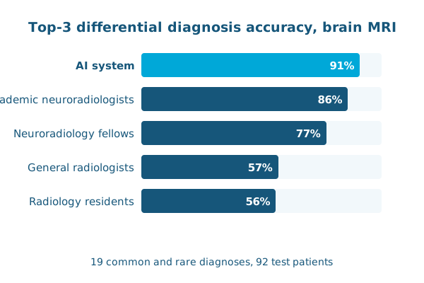
Combining deep learning with Bayesian networks, an AI system matched academic neuroradiologists at differential diagnosis for 19 diseases on brain MRI. Published in Radiology.
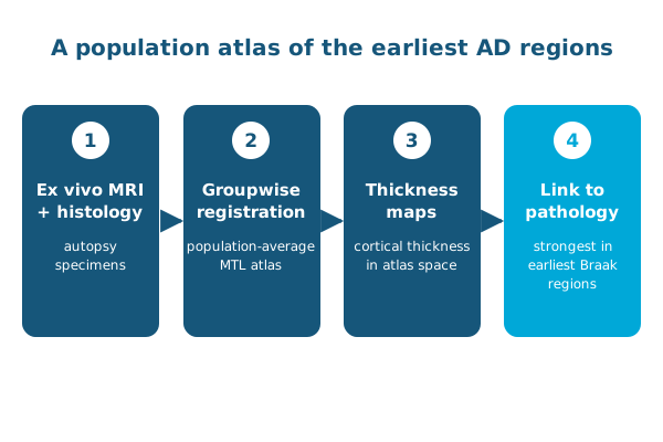
Groupwise registration of ex vivo MRI builds a population atlas of the medial temporal lobe that links cortical thinning to Alzheimer’s pathology in the earliest Braak regions. Presented at ISBI 2020.
2019
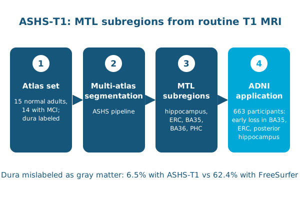
A new atlas set that labels the dura lets ASHS segment MTL subregions on routine T1 MRI, detecting early atrophy in prodromal Alzheimer’s disease. Published in Human Brain Mapping.
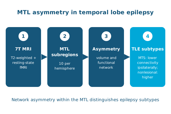
7T structural and functional MRI shows different patterns of MTL asymmetry in lesional and MRI-negative temporal lobe epilepsy. Published in Human Brain Mapping.
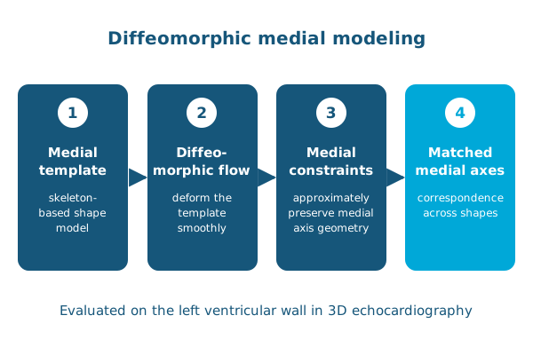
A shape analysis approach that combines medial modeling with diffeomorphic flows, keeping skeletons in correspondence as shapes deform. Presented at IPMI 2019.
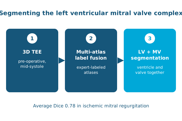
Multi-atlas label fusion segments the left ventricular mitral valve complex in pre-operative 3D TEE, a step toward predicting outcomes in ischemic mitral regurgitation. Presented at STACOM.
2018
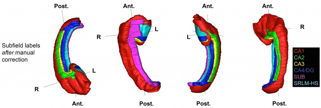
Ex vivo MRI of 31 hippocampal specimens, labeled using histology, yields a 3D probabilistic atlas of hippocampal subfields and their changes in aging and Alzheimer’s disease. Published in PNAS.
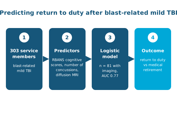
In 303 service members with blast-related mild TBI, cognitive testing and concussion history predicted return to duty versus medical retirement. Published in the Journal of Neurotrauma.
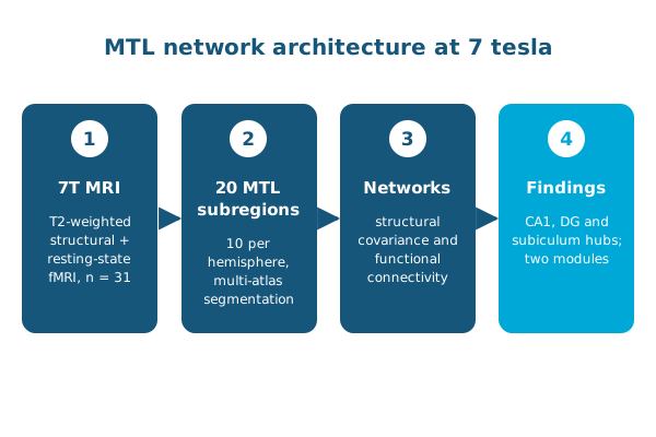
Structural and functional 7 tesla MRI of 31 healthy adults reveals hub subregions and a two-module network organization in the medial temporal lobe. Published in Human Brain Mapping.
2017
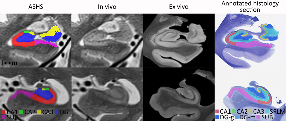
Registering 3T in vivo and 9.4T ex vivo scans of the same individuals shows how to validate in vivo hippocampal subfield segmentation against tissue. Published in Cerebral Cortex.
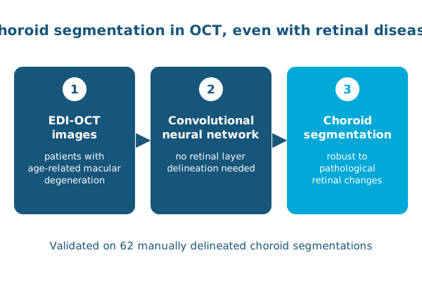
A convolutional neural network segments the choroid in OCT images of patients with macular degeneration without first finding the retinal layers. Presented at the OMIA workshop at MICCAI 2017.
2016

We recently examined potential relationship between tau protein uptake in medial temporal lobe (MTL) and volumetric measures of MTL subregions in a small dataset of Alzheimer’s disease patients. Since tau protein…
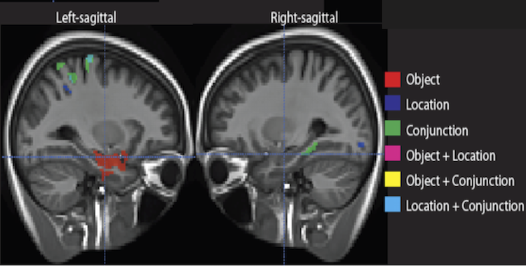
Medial temporal lobe (MTL) is generally thought to be a principal site of long term memory (LTM) process, and isn’t usually associated with short term memory (STM). Some recent work have suggested a role for MTL in STM…
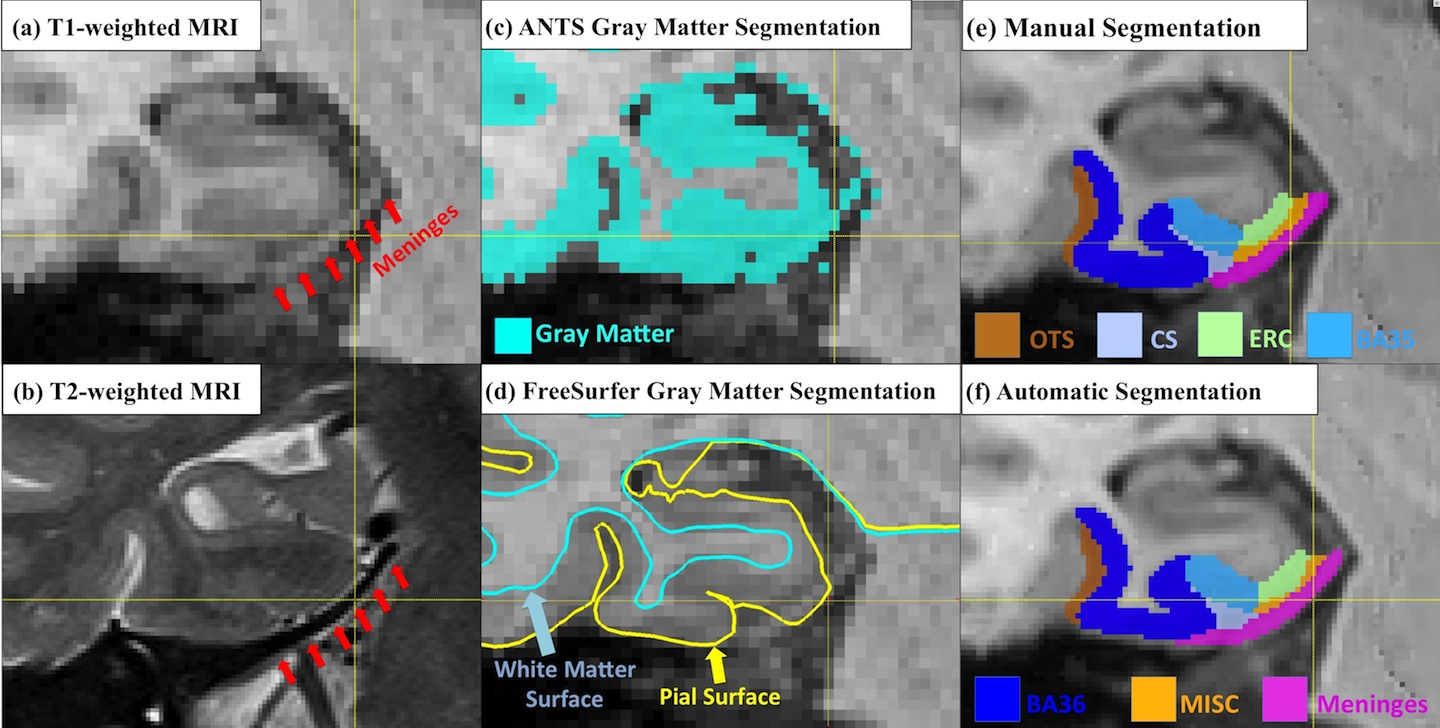
Quantification of medial temporal lobe (MTL) cortices, including entorhinal cortex (ERC) and perirhinal cortex (PRC), from in vivo MRI is desirable for studying the human memory system as well as in early diagnosis and…
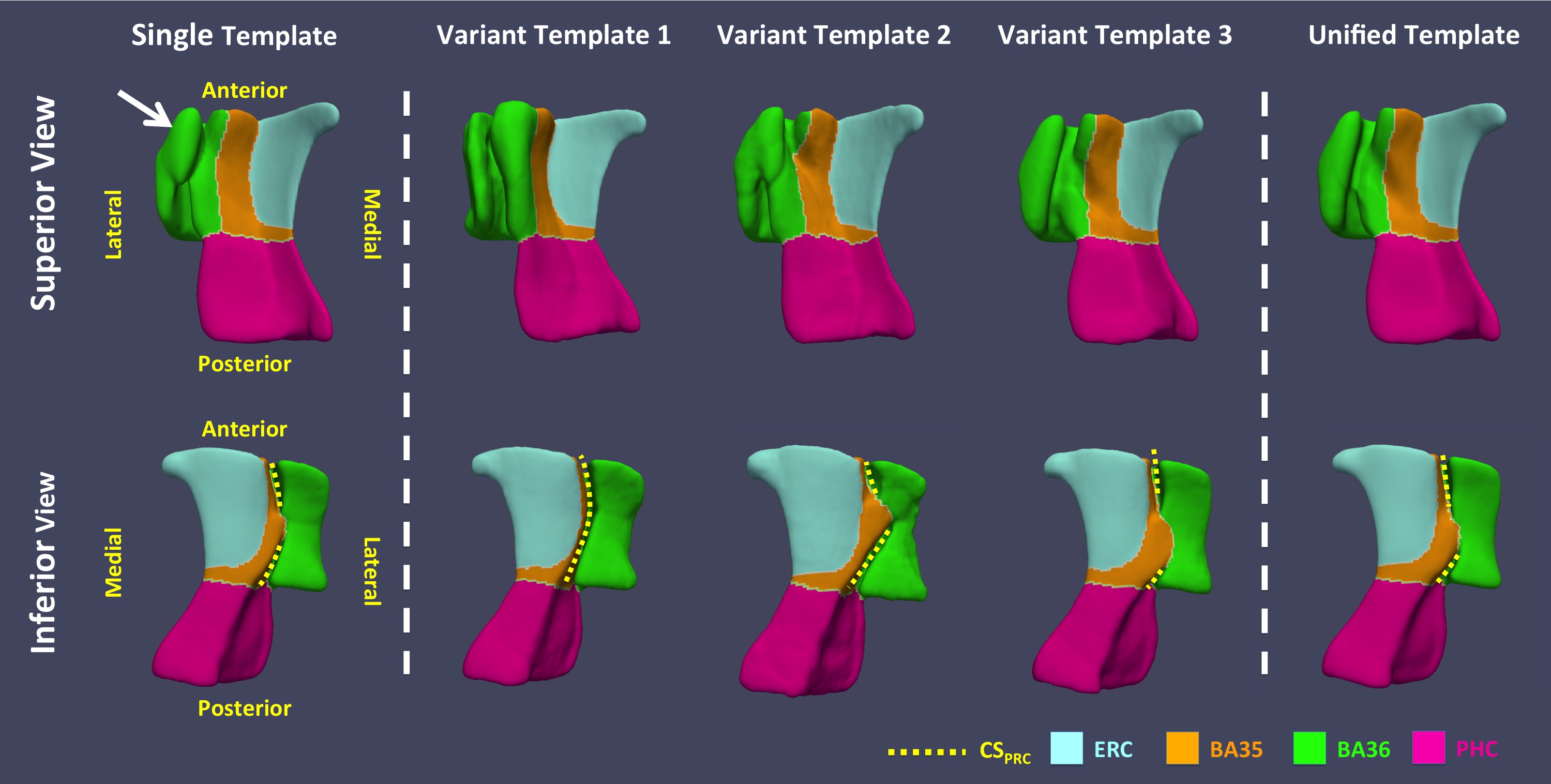
The human perirhinal cortex (PRC) plays critical roles in episodic and semantic memory and visual perception. The PRC consists of Brodmann areas 35 and 36 (BA35, BA36). In Alzheimer’s disease (AD), BA35 is the first…
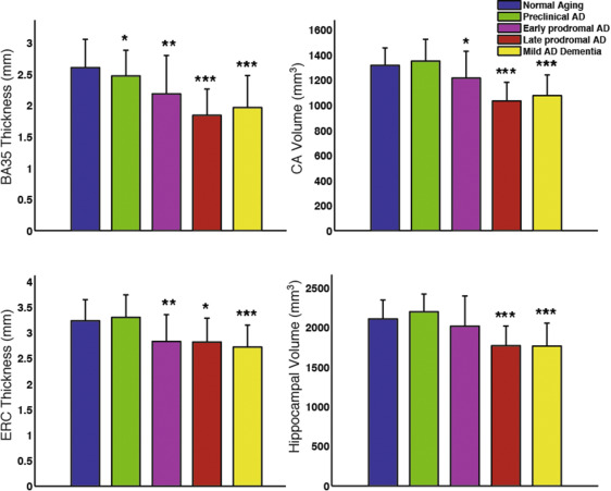
Group comparison of select MTL subregional measurements adjusted for age and ICV. Statistical significance is based on comparison with the “normal aging” group (amyloid-negative ADNI controls). Error bars reflect 1…
In our recent publication in Cerebral Cortex we compared in vivo and ex vivo MRI scans of the human hippocampus and evaluated to ability to match information between the two modalities. We were able to validate several…
2015
Our paper showing that psychometric tests predict Alzheimer’s Disease pathology in Mild Cognitive Impairment subjects was just accepted by the Journal of Alzheimer’s Disease. Read the in-press version here.
2014
Our paper on multi-modal dimensionality reduction was recently accepted by Methods. It shows how to use our sparse dimensionality reduction techniques to form interpretable predictive models from neuroimaging data. All…
We designed a method to construct structural cortical graphs in a manner similar to BOLD-based functional connectivity graphs (paper, poster at MICCAI 2014).
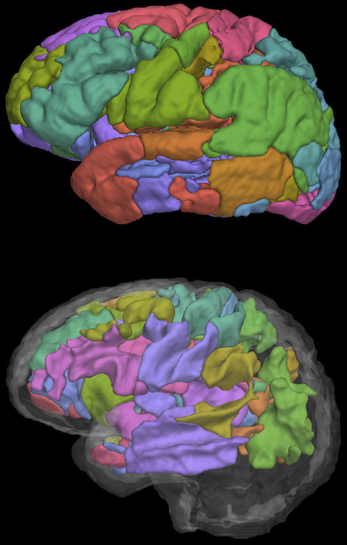
Linking structural neuroimaging data from multiple modalities to cognitive performance is an important challenge for cognitive neuroscience. In this study we examined the relationship between verbal fluency performance…
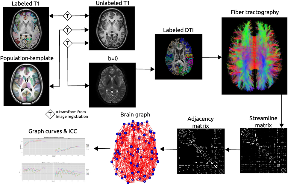
Recent interest in human brain connectivity has led to the application of graph theoretical analysis to human brain structural networks, in particular white matter connectivity inferred from diffusion imaging and fiber…
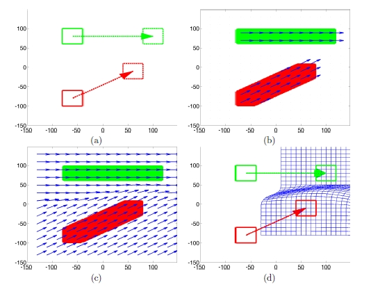
We propose a novel way to construct a diffeomorphic polyaffine transformation model. Each affine transform is defined on a local region and the resulting diffeomorphism encapsulates all the local transforms by a smooth…
Interactive image registration is required by some medical applications since automatic image registration is often slow and sometimes error-prone. We consider interactive registration methods that incorporate…
Extra-hippocampal medial temporal lobe (ehMTL) structures, which consists of entorhinal (ERC) and perirhinal (PRC) cortices, receives increasing attention because they are amongst the earliest sites affected by…
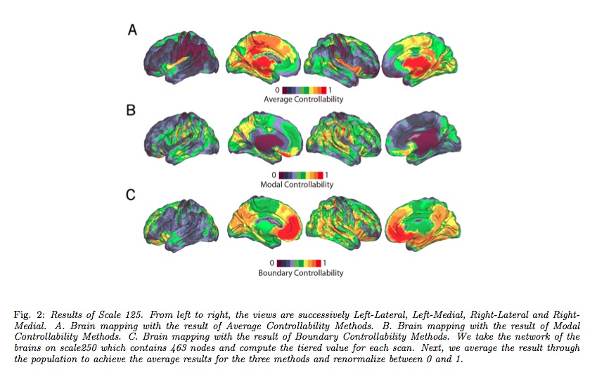
Neuroscientific investigations seek to reveal how neural systems perform complex functions, how those systems and functions are altered in disease states, and how therapeutic interventions can be used to redirect these…
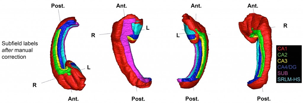
We are constructing a computational, probabilistic atlas of the human hippocampal formation (HF) from postmortem, high-resolution 9.4T MRI and densely acquired histology. The human HF consists of several structures of…
We have recently updated ANTs to perform processing of perfusion ASL images. The pipeline is designed to be consistent with the antsCorticalThickness pipeline for computing cortical thickness and incorporates…
The complex and high-dimensional nature of medical images makes them difficult to incorporate into traditional linear model-based statistical analysis. One way of simplifying analysis of medical images is to use a…
Our paper on sparse regression for predicting cognitive performance from cortical thickness was published at Information Processing in Medical Imaging (IPMI) 2013. We developed a method for performing regression on…
We have been developing a framework to incorporate ITK and ANTs-based image processing methods into the R programming language to allow for interactive statistical analysis of medical images. R provides the most…
Our paper looking at dissociable functional networks of anterior and posterior MTL regions in older healthy controls and MCI patients just got accepted for publication in Neurobiology of Aging. We labeled MTL subregions…
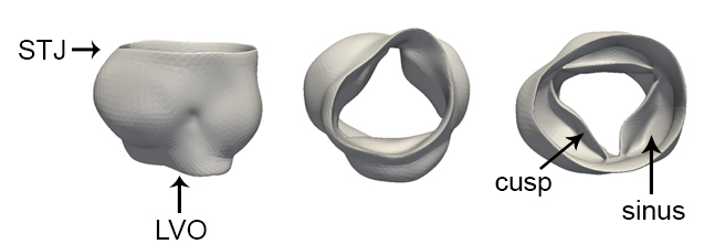
Given the importance of image-based morphological assessment in the diagnosis and surgical treatment of aortic valve disease, there is considerable need to develop a standardized framework for 3D valve segmentation and…
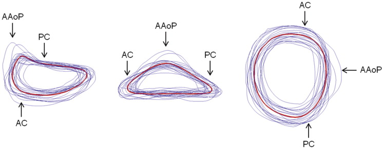
The basis of mitral annuloplasty ring design has progressed from qualitative surgical intuition to experimental and theoretical analysis of annular geometry with quantitative imaging techniques. In this work, we present…
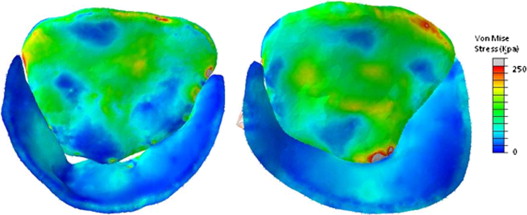
An integrated methodology for imaging, segmenting, modeling, and deriving computationally-predicted pressure-derived mitral leaflet stresses is presented and points the way towards intraoperative and periprocedural…
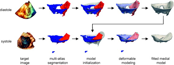
Comprehensive visual and quantitative analysis of in vivo human mitral valve morphology is central to the diagnosis and surgical treatment of mitral valve disease. Real-time 3D transesophageal echocardiography (3D TEE)…
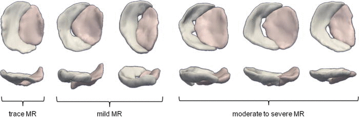
Precise 3D modeling of the mitral valve has the potential to improve our understanding of valve morphology, particularly in the setting of mitral regurgitation (MR). Toward this goal, we have developed a…
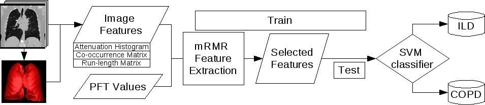
To compare the performance of various image-based metrics computed from thoracic HRCT modality with data from pulmonary function testing (PFT) in characterizing interstitial lung disease (ILD) and chronic obstructive…
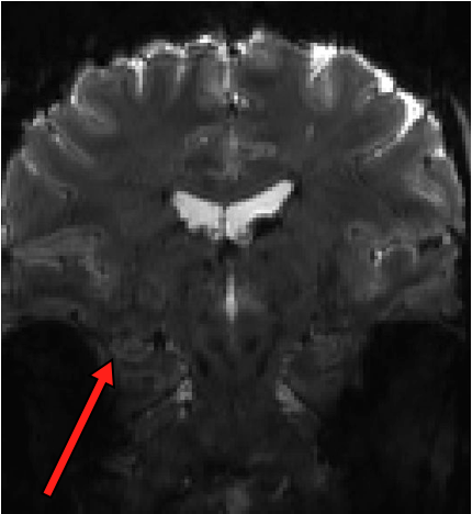
We just found out that our R03 grant application for high-resolution BOLD fMRI at 7T has been recommended for funding. We have been anxiously waiting for the funding decision which was delayed by the government shutdown…
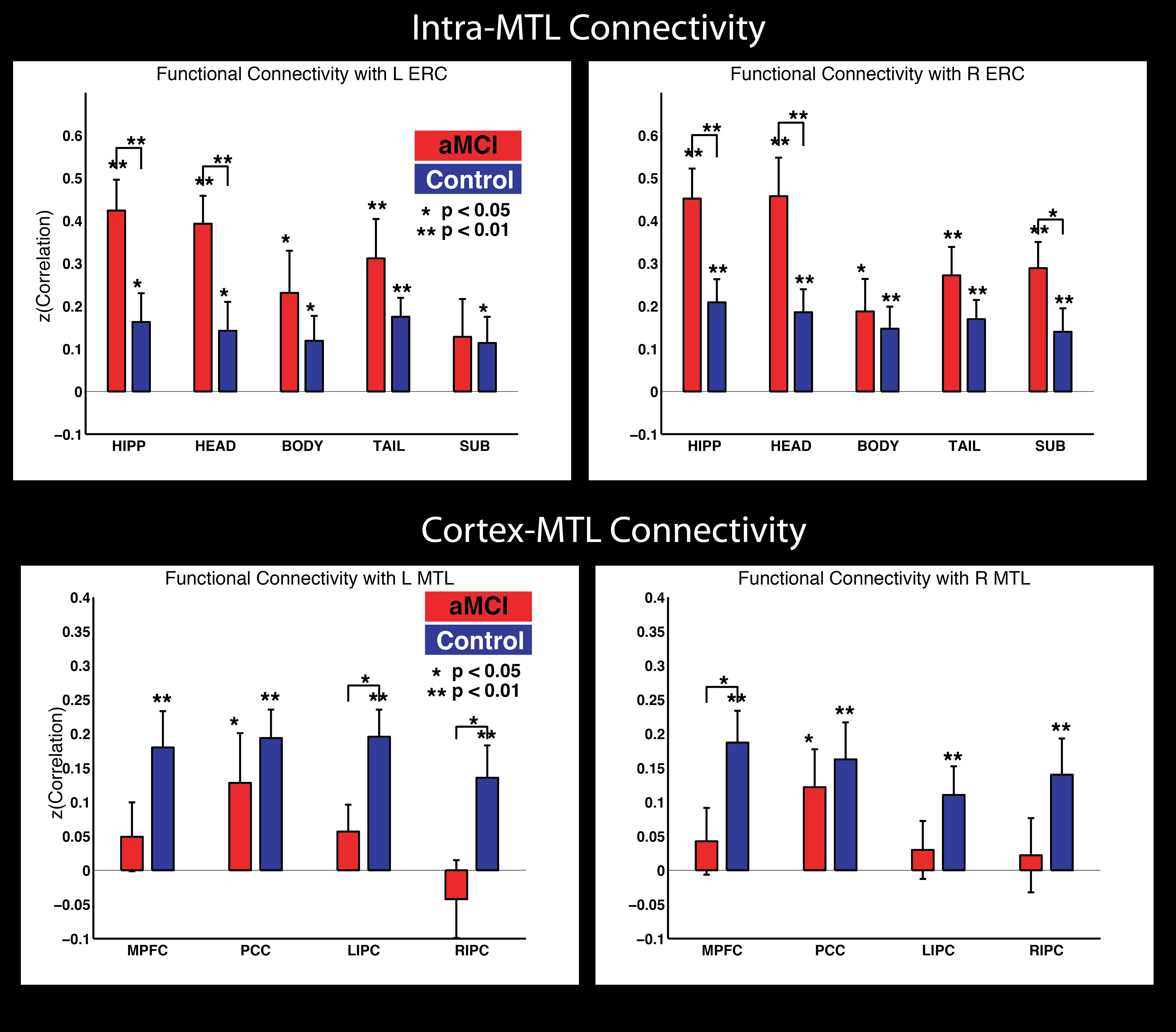
There is a ton of work looking at BOLD functional connectivity (FC) in Alzheimer’s Disease, and one consensus that has emerged is that this is a disconnection syndrome, with most studies finding weaker connectivity…
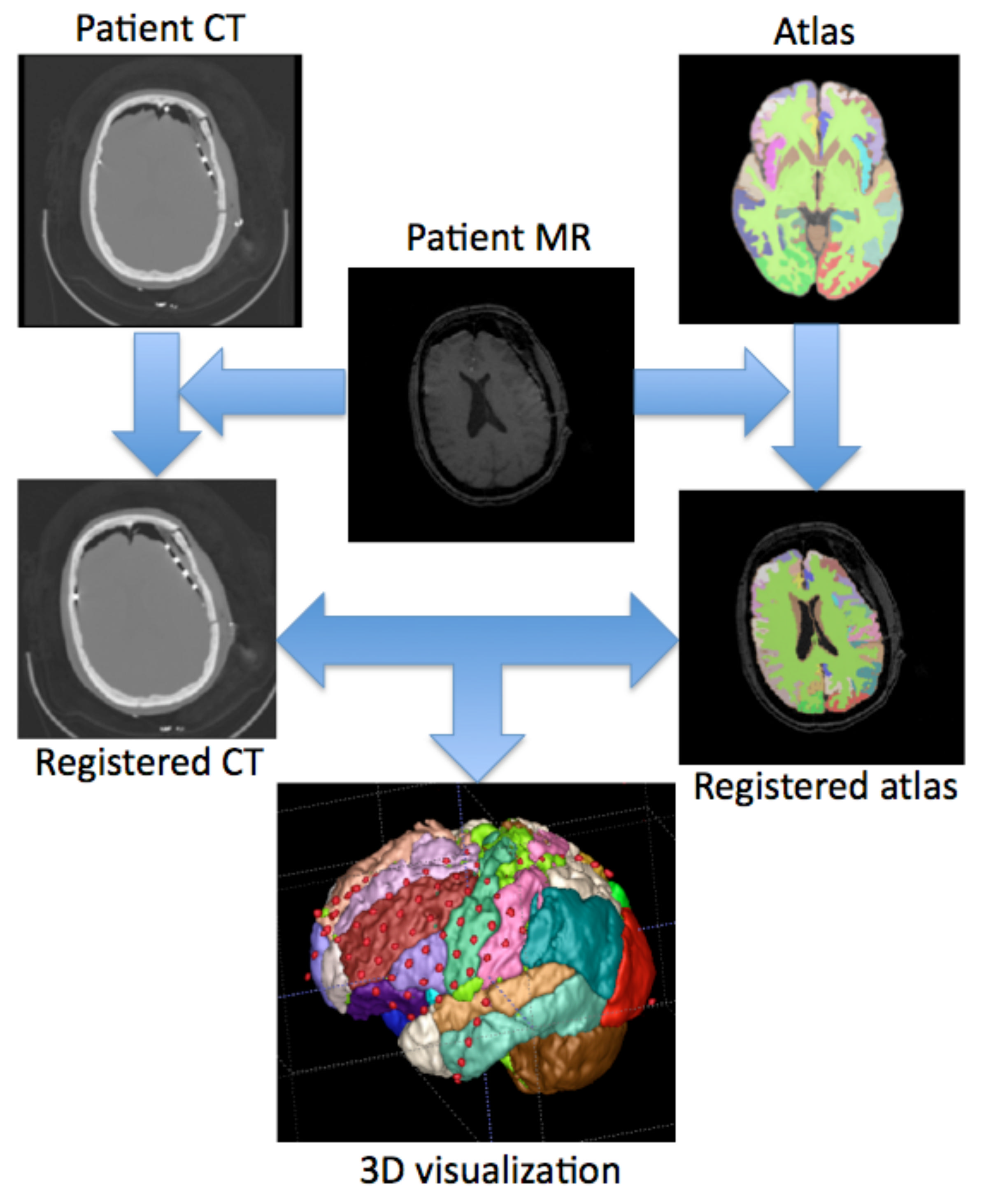
Objective: Visualizing implanted subdural electrodes in 3D space can greatly aid planning, executing, and validating resection in epilepsy surgery. Co-registration software is available, but cost, complexity,…
In the absence of a neonatal template with cortical subregion labels, it can be extremely difficult to obtain cortical parcellation of new neonatal brain images automatically. We address this problem by utilizing adult…
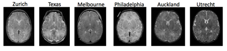
The International Consortium on CHD Neonatal Brain MRI was established to coordinate efforts to tackle this problem of MR data normalization and provide an open common platform with tools for processing and analyzing…
This research seeks to understand the relations among socioeconomic status (SES), early life experience, and learning in adolescents. Previous research has shown that learning ability is positively correlated with SES,…
Children growing up in poverty are at increased risk for poor cognitive outcomes, arguably one of the most critical issues facing our nation today. While many studies have shown that effects of poverty are apparent by…
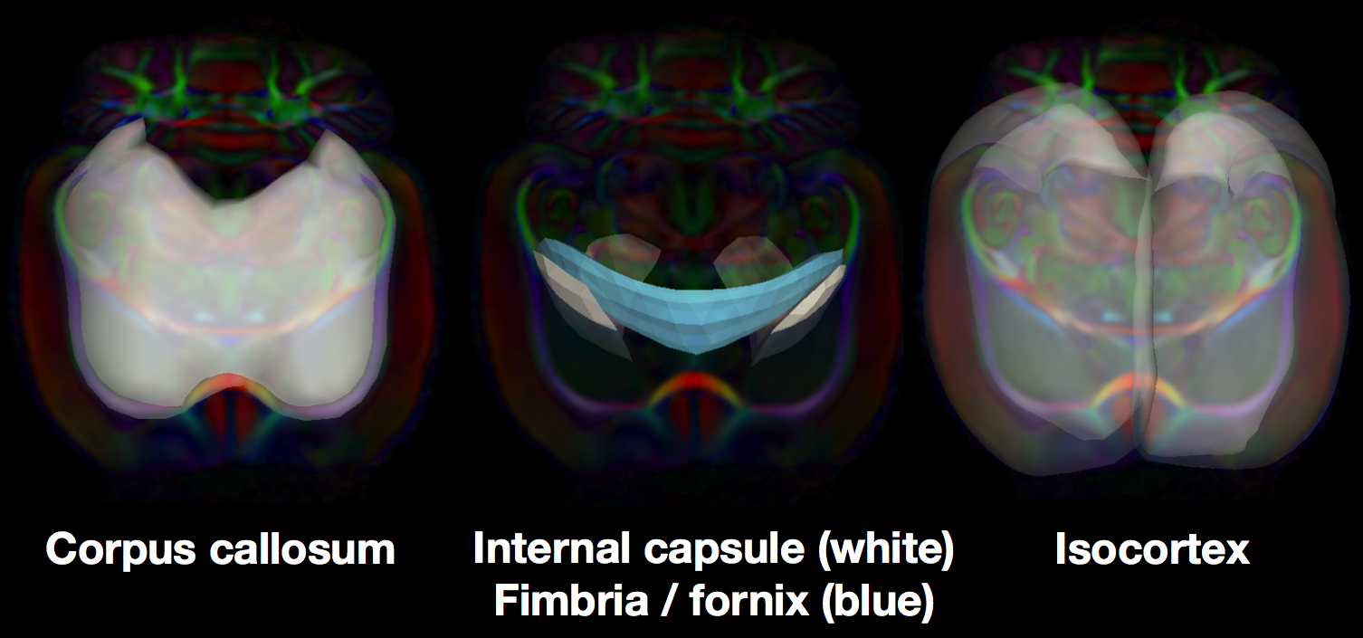
Tract specific Analysis (TSA) uses continuous medial modeling for structure- specific analysis of diffusion in sheet-like white matter tracts in the human brain. The medial geometry provides both a skeleton surface (the…
2013
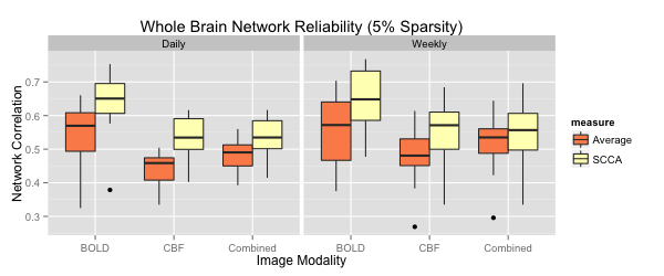
We contribute a novel multivariate strategy for computing the structure of functional networks in the brain from arterial spin labeling (ASL) MRI. Our method fuses and correlates multiple functional signals by employing…
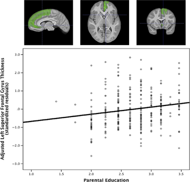
Childhood socioeconomic status (SES) predicts executive function performance and measures of prefrontal cortical function, but little is known about its anatomical correlates. Structural MRI and demographic data from a…
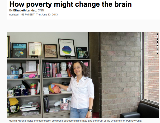
Across the Penn campus in the radiology department, Farah sits in a low chair while Brian Avants, assistant professor of radiology, explains their recent study, using a slide on a computer screen. Farah presented the…
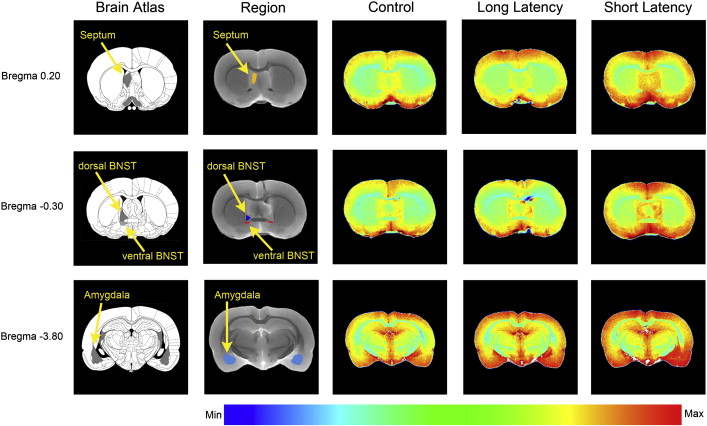
Swim stress produces relatively different magnitudes of Mn2 + enhancement in certain brain regions in rats with different coping strategies. The panels illustrate differences in Mn2 + enhancement in the septum, BNST,…
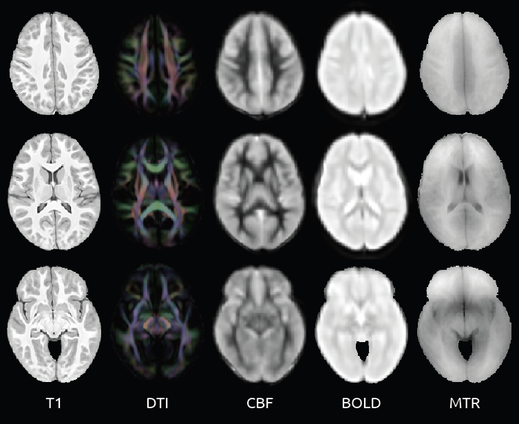
Sample slices from the multivariate atlas used as a basis for neuro-anatomical comparison [Could not find the bibliography file(s) Purpose: To evaluate the relationships between cerebral blood flow and other magnetic…
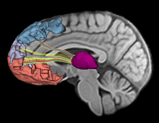
This collaboration with the Moss Rehabilitation Research Institute at the Albert Einstein Healthcare Network involves the development of methologies for examining both structural and connective properties in the brains…
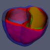
The aims of this collaborative project with the Computational Imaging Lab at the Pompeu Fabra University are to use a medial model of the myocardium to generate stronger shape priors for segmentation, richer features…
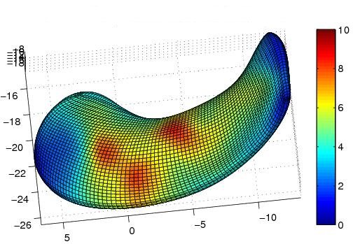
We are developing a new class of techniques that focus statistical analysis of functional neuroimaging data on specific structures such as the hippocampus, using the shape of the structures as the guide by which to…
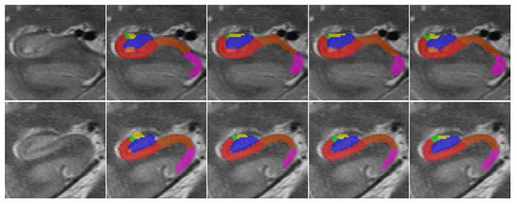
Segmentation, the problem of locating and outlining objects of interest in images, is a central problem in biomedical image analysis. It is the primary mechanism for quantifying the properties of anatomical structures…
Research areas
Using MRI to examine brain function
Traumatic Brain Injury (TBI) Post Traumatic Stress Disorder (PTSD)