
3D glutamate imaging at 7T locates seizure onset in nonlesional epilepsy

In about a third of patients with drug-resistant epilepsy, standard MRI shows no lesion, and these patients have worse outcomes after surgery. Earlier work showed that glutamate imaging with GluCEST (glutamate chemical exchange saturation transfer) could identify the side of seizure onset in nonlesional temporal lobe epilepsy (TLE), but only in a single 2D slice.
In Brain and Behavior, Peter Hadar, Laura Wisse, Sandhitsu Das, Kathryn Davis and colleagues extended GluCEST to three dimensions on a 7 tesla scanner. The analysis used the ANTs pipeline and the ASHS hippocampal subfield segmentation.
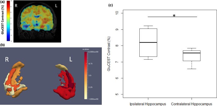
In four MRI-negative TLE patients with left-sided seizure onset, volumetric GluCEST showed higher glutamate in the hippocampus on the side of seizure onset, and also in the subiculum on that side. Localizing seizure onset down to a hippocampal subfield is an improvement on the earlier single-slice method, and points toward whole-brain, noninvasive imaging of glutamate networks in epilepsy.
Reference: Hadar PN, Kini LG, Nanga RPR, et al., Das S, Davis KA. Volumetric glutamate imaging (GluCEST) using 7T MRI can lateralize nonlesional temporal lobe epilepsy: a preliminary study. Brain and Behavior. 2021. doi:10.1002/brb3.2134