
A 3D atlas of the human hippocampus from ex vivo MRI and histology

The hippocampus is one of the most studied structures in the human brain, yet little quantitative data exist on its 3D organization, how much it varies between people, and how disease affects its subregions. Histology provides only limited reference information because it is two-dimensional.
In a study in the Proceedings of the National Academy of Sciences, Daniel Adler, Paul Yushkevich and colleagues combined very high resolution (about 200 μm) ex vivo MRI scans of 31 human hippocampal specimens into a 3D probabilistic atlas, using groupwise diffeomorphic registration. Serial histology in 9 of the specimens was used to label the hippocampal subfields in the atlas based on cytoarchitecture. The specimens came from donors with Alzheimer’s disease (AD), other dementias, and no dementia.
Analyzing the specimens in atlas space showed:
- Disproportionate involvement of the CA1 subfield and the stratum radiatum lacunosum moleculare in AD, with lesser involvement of the dentate gyrus and CA2/3.
- An association with age in the dentate gyrus and, to a lesser extent, CA1.
These 3D patterns of variability, disease and aging effects are directly relevant to the growing field of hippocampal subfield imaging in living people.
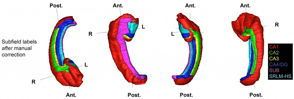
Other PICSL contributors include Laura Wisse, Ranjit Ittyerah, Long Xie, Jiancong Wang and Sandhitsu Das.
Reference: Adler DH, Wisse LEM, Ittyerah R, et al., Yushkevich PA. Characterizing the human hippocampus in aging and Alzheimer’s disease using a computational atlas derived from ex vivo MRI and histology. Proceedings of the National Academy of Sciences. 2018. doi:10.1073/pnas.1801093115