
Deep learning measures of tau and TDP-43 explain medial temporal lobe atrophy

The medial temporal lobe (MTL) is a hotspot for neuropathology, and MTL atrophy is widely used as a biomarker of cognitive decline. Because several proteins accumulate there at once, it has been hard to say which pathology drives the atrophy.
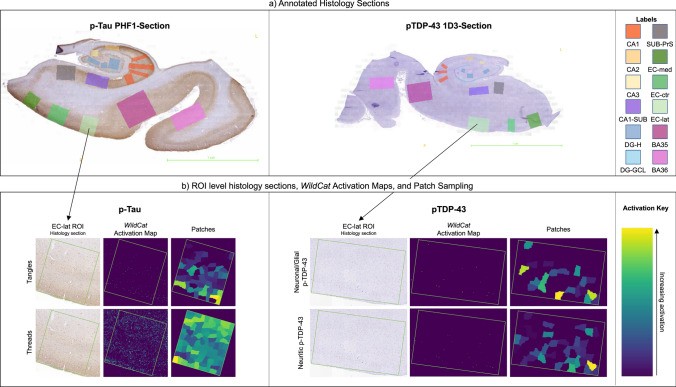
In a study in Acta Neuropathologica, Amanda Denning, Paul Yushkevich and colleagues developed two deep learning algorithms that measure phosphorylated tau (p-tau) and TDP-43 pathology on digitized histology sections with annotated MTL subregions. The focus was on Alzheimer’s disease and limbic-predominant age-related TDP-43 encephalopathy (LATE).
Key findings:
- Both quantitative measures agree closely with expert visual ratings and distinguish well between pathology stages.
- In 140 cases with MRI scans taken during life, p-tau pathology was associated with structure across MTL subregions, while TDP-43 had more limited associations with the hippocampus and entorhinal cortex.
- Quantitative p-tau measures explained antemortem cortical thickness and volume significantly better than semi-quantitative ratings, and still showed associations in severe AD cases where the ratings hit a ceiling.
The results argue for quantitative neuropathology in future studies of how pathology relates to brain structure.
Other PICSL contributors include Ranjit Ittyerah, Lisa Levorse, Chinmayee Athalye, Sadhana Ravikumar, Mengjin Dong, Yue Li, Pulkit Khandelwal, Laura Wisse and Sandhitsu Das.
Reference: Denning AE, Ittyerah R, Levorse LM, et al., Yushkevich PA. Association of quantitative histopathology measurements with antemortem medial temporal lobe cortical thickness in the Alzheimer’s disease continuum. Acta Neuropathologica. 2024. doi:10.1007/s00401-024-02789-9