
High-resolution imaging of the medial temporal lobe in ADNI

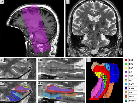
Since Phase 3, the Alzheimer’s Disease Neuroimaging Initiative (ADNI) has included a high-resolution T2-weighted MRI scan optimized for imaging hippocampal subfields and the subregions of the medial temporal lobe (MTL), the part of the brain where Alzheimer’s disease first takes hold. These scans are processed by the ADNI core to produce automatic segmentations and morphological measurements.
In a paper for ADNI’s 20th anniversary in Alzheimer’s & Dementia, Paul Yushkevich, Sandhitsu Das and colleagues review:
- Why: the work that supported adding this imaging modality to ADNI3.
- How: the ADNI MTL imaging and analysis protocols, and a summary of the studies that have used the data.
- What’s next: an outlook for high-resolution MTL imaging in ADNI Phase 4.
The review includes a new study that uses surface-based tools to characterize MTL neurodegeneration across biomarker-defined AD stages. It reveals a spreading pattern of cortical thinning that grows with tau pathology in the presence of elevated amyloid, with apparent epicenters in the transentorhinal region and the inferior hippocampal subfields. This closely matches postmortem descriptions of how tau spreads, supporting tau, in the presence of amyloid, as the driver of neurodegeneration.
Other PICSL contributors include Ranjit Ittyerah, Yue Li, Amanda Denning, Sydney Lim, Emily McGrew, Long Xie, Robin de Flores and Laura Wisse.
Reference: Yushkevich PA, Ittyerah R, Li Y, et al., Wolk DA, Das SR. Morphometry of medial temporal lobe subregions using high-resolution T2-weighted MRI in ADNI3: why, how, and what’s next? Alzheimer’s & Dementia. 2024. doi:10.1002/alz.14161