
Tau pathology and cortical thinning in whole-hemisphere 7T MRI

Neurodegenerative diseases involve several pathologies that often occur together, but none of them can be measured specifically in living patients. Linking patterns of tissue loss on MRI to the pathologies underneath could turn structural MRI into a probabilistic biomarker.
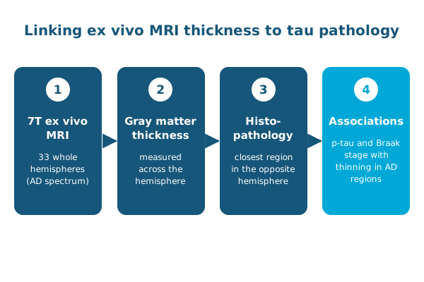
In Alzheimer’s & Dementia, Shokufeh Sadaghiani, Paul Yushkevich and colleagues imaged 33 whole brain hemispheres from donors on the Alzheimer’s disease (AD) spectrum with T2-weighted 7 tesla MRI. They paired gray matter thickness with histopathology from the closest anatomical region in the opposite hemisphere.
- Accounting for TDP-43 and α-synuclein, age, sex and postmortem interval, phosphorylated tau was related to cortical thickness in the entorhinal and primary visual cortices, temporal pole, insula and posterior cingulate.
- Braak stage was correlated with thickness in the parahippocampal gyrus, entorhinal cortex and Brodmann area 35.
Even with a limited range of pathology in these cases, measures of AD pathology were associated with tissue loss in several AD regions, which helps interpret structural MRI studies in living patients.
Other PICSL contributors include Sydney Lim, Ranjit Ittyerah, Sadhana Ravikumar, Pulkit Khandelwal, Sandhitsu Das and Laura Wisse.
Reference: Sadaghiani S, Trotman W, Lim SA, et al., Yushkevich PA. Associations of phosphorylated tau pathology with whole-hemisphere ex vivo morphometry in 7 tesla MRI. Alzheimer’s & Dementia. 2023 (published online 2022). doi:10.1002/alz.12884