
Medial temporal lobe atrophy patterns in semantic variant PPA

Semantic variant primary progressive aphasia (svPPA) is a dementia in which people progressively lose the meaning of words and objects. Studies with T1 MRI have shown early atrophy in the left anterior temporal lobe, especially the perirhinal cortex. High-resolution T2 MRI now makes it possible to measure the subregions of the medial temporal lobe (MTL) in much finer detail.
In Neurobiology of Aging, Laura Wisse, Ranjit Ittyerah, Sydney Lim, Paul Yushkevich, Sandhitsu Das, Murray Grossman and colleagues measured MTL subregions with ASHS in 15 patients with svPPA, 37 with Alzheimer’s disease (AD) and 23 healthy controls. A subset was followed over time with deformation-based morphometry.
- Compared with controls, svPPA patients had volume loss in the left MTL, right CA1, and Brodmann areas 35 and 36 (subdivisions of the perirhinal cortex).
- Compared with AD patients, they had smaller left CA1 and BA35, and smaller BA36 on both sides.
- Over time, BA36 atrophied faster in svPPA than in controls.
Fine-grained MTL measures could help track disease progression in svPPA and serve as markers in clinical trials.
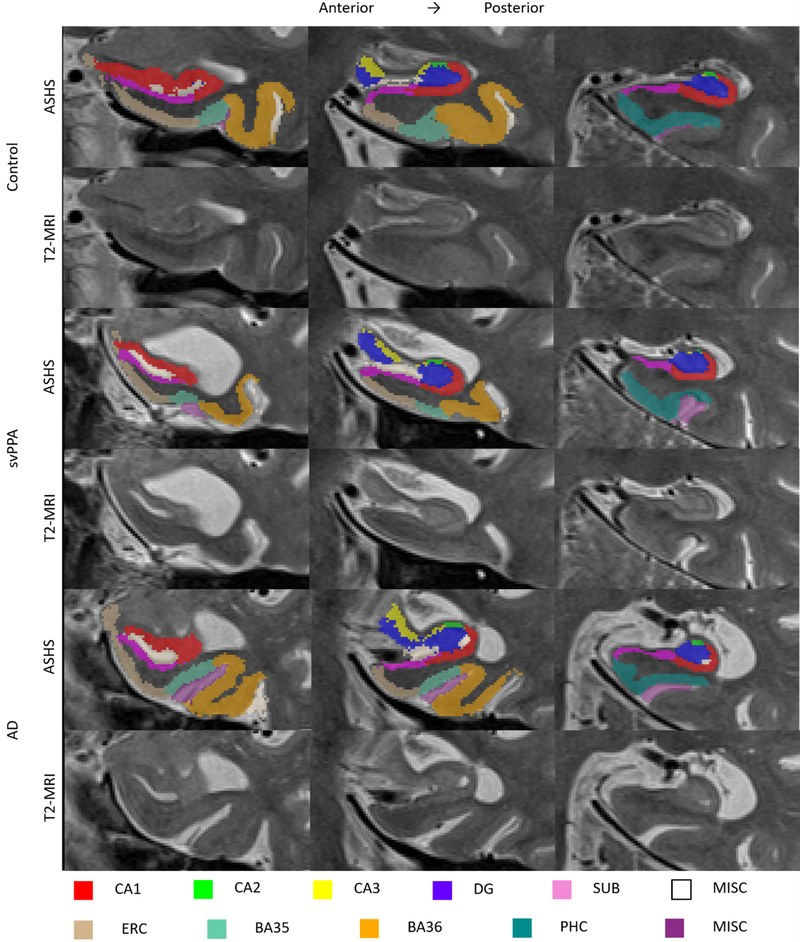
Reference: Wisse LEM, Ungrady MB, Ittyerah R, et al., Das SR, Grossman M. Cross-sectional and longitudinal medial temporal lobe subregional atrophy patterns in semantic variant primary progressive aphasia. Neurobiology of Aging. 2021. doi:10.1016/j.neurobiolaging.2020.11.012