
Postmortem MRI links deep brain atrophy to specific neurodegenerative pathologies

Alzheimer’s disease and related dementias are caused by different misfolded proteins, including tau, α-synuclein and TDP-43, and many patients carry more than one. How each of these pathologies affects deep brain structures such as the hippocampus, amygdala, thalamus and basal ganglia is still poorly understood, largely because most of these pathologies cannot be measured directly in living patients.
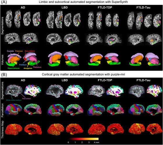
In a study published in Alzheimer’s & Dementia, Pulkit Khandelwal, Paul Yushkevich and colleagues across Penn combined high-resolution postmortem MRI with detailed neuropathology from 132 brain donors. The donors had Alzheimer’s disease (AD), Lewy body disease (LBD), frontotemporal lobar degeneration with TDP-43 (FTLD-TDP), or FTLD with tau. Automated deep learning tools segmented seven subcortical and limbic structures and reconstructed the cortical surface of each scanned hemisphere, so structure volumes could be related to cortical thickness and to measured pathology.
Key findings:
- LBD showed the least subcortical, limbic and cortical atrophy of the four groups. In donors with both AD and LBD pathology, primary LBD was associated with less atrophy than primary AD.
- Each disease had its own pattern of coupling between deep structures and the cortex: AD showed broad cortico-subcortical and cortico-limbic associations, while LBD’s were limited to parieto-occipital cortex.
- In AD and FTLD-tau, hippocampal volume tracked p-tau burden, neuronal loss and gliosis. In LBD, the severity of α-synuclein in the thalamus was associated with subcortical and limbic volumes.
Together, these results show that postmortem neuroimaging can reveal disease- and region-specific links between brain structure and pathology. That knowledge is needed to interpret atrophy measured in living patients.
PICSL and PATCH Lab contributors include Lisa Levorse, Sydney Lim, Amanda Denning, Christopher Olm, Ranjit Ittyerah, Laura Wisse and Sandhitsu Das.
Reference: Khandelwal P, Duong MT, Levorse LM, et al., Yushkevich PA. Postmortem brain MRI reveals differential associations of subcortical and limbic volumes with cortical thinning and neurodegenerative pathologies. Alzheimer’s & Dementia. 2026. doi:10.1002/alz.71649