
Medial temporal lobe asymmetry in temporal lobe epilepsy at 7T

Mesial temporal lobe epilepsy (TLE) is a common neurological disorder affecting the hippocampus and surrounding medial temporal lobe (MTL). Whole-brain network changes in TLE had been studied, but not the functional network of the MTL at the level of its subregions.
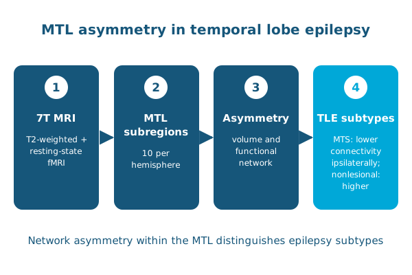
In Human Brain Mapping, Preya Shah, Laura Wisse, Paul Yushkevich, Sandhitsu Das, Kathryn Davis and colleagues used high-resolution 7T T2-weighted MRI and resting-state fMRI in patients with unilateral, drug-resistant TLE and in healthy controls. Patients were split into those with mesial temporal sclerosis (TLE-MTS) and those whose MRI showed no lesion (TLE-NL). An automated multi-atlas pipeline delineated 10 MTL subregions per hemisphere.
- In volume, TLE-MTS patients had smaller hippocampal subfields on the side of seizure onset, while TLE-NL patients showed only a mild decrease in whole-hippocampal volume.
- In function, the CA1 subfield and whole hippocampus showed opposite asymmetries: increased connectivity on the seizure side in TLE-NL, and decreased connectivity in TLE-MTS.
Network properties within the MTL can therefore distinguish TLE subtypes, and high-resolution MRI could improve localization for patients being considered for surgery.
Reference: Shah P, Bassett DS, Wisse LEM, et al., Das SR, Davis KA. Structural and functional asymmetry of medial temporal subregions in unilateral temporal lobe epilepsy: a 7T MRI study. Human Brain Mapping. 2019. doi:10.1002/hbm.24530