
A simple hippocampal volume cut-off flags likely LATE co-pathology

Limbic-predominant age-related TDP-43 encephalopathy (LATE) often occurs together with Alzheimer’s disease (AD) and causes similar symptoms. With no validated in vivo biomarker for LATE, identifying patients with mixed AD/LATE pathology is difficult, yet it matters for prognosis and for clinical trials.
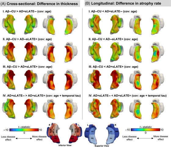
In a study in Alzheimer’s & Dementia, Nidhi Mundada, Sandhitsu Das, David Wolk and colleagues tested a simple approach. Among cognitively impaired ADNI participants with MRI, amyloid PET and tau PET, people in the lowest quartile of hippocampal volume were labeled as having suspected LATE (sLATE). Combined with amyloid status, this defined three groups: AD+sLATE−, AD−sLATE+ and AD+sLATE+.
Key findings:
- The AD+sLATE+ group showed greater atrophy of the anterior hippocampus and amygdala than AD+sLATE−.
- Both sLATE+ groups showed greater anterior MTL atrophy and worse memory and language performance.
- AD+sLATE+ also showed faster cognitive decline.
- The classification was validated in an autopsy cohort.
Because it requires no complex modeling or biomarker panels, this cut-off is a practical, scalable way to flag likely mixed pathology and to enrich or exclude such patients in clinical trials.
Other PICSL contributors include Xueying Lyu, Emily McGrew, Long Xie, Philip Cook, Jim Gee and Paul Yushkevich.
Reference: Mundada NS, Lyu X, Brown CA, et al., Yushkevich PA, Das SR, Wolk DA. Enrichment of patients with concomitant limbic-predominant age-related TDP-43 encephalopathy (LATE) on the Alzheimer’s disease continuum using hippocampal volume. Alzheimer’s & Dementia. 2025. doi:10.1002/alz.70970