
An open-source tool for designing synthetic skeletons

Deformable medial modeling represents anatomy by its skeleton, an approach that supports statistical shape analysis and patient-specific measurements such as locally varying thickness. It works by deforming a predefined synthetic skeleton, or template, to fit each anatomical structure. Until now, the lack of software for creating these skeletons has limited the method’s wider use.
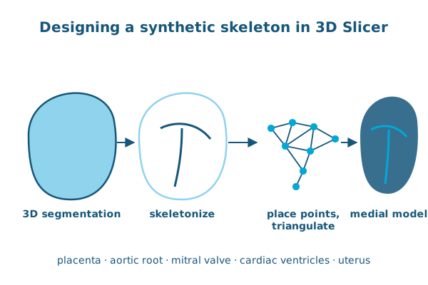
In the Journal of Medical Imaging, Christian Herz, Abdullah Aly, Paul Yushkevich, Alison Pouch and colleagues present an open-source user interface for designing synthetic skeletons, built into 3D Slicer. A user imports and skeletonizes a 3D segmentation, then interactively places points in 3D and triangulates the medial surface to get the desired branching structure.
Three users designed skeletons for anatomies with a range of topologies: the placenta, aortic root wall, mitral valve, cardiac ventricles and uterus. The skeletons worked with both skeleton-first and boundary-first medial modeling software, and the fitted models represented the original segmentations accurately.
Reference: Herz C, Vergnet N, Tian S, et al., Yushkevich PA, Pouch AM. Open-source graphical user interface for the creation of synthetic skeletons for medical image analysis. Journal of Medical Imaging. 2024. doi:10.1117/1.JMI.11.3.036001