
A 3D atlas of tau tangles in the medial temporal lobe

Tau neurofibrillary tangles (NFTs) first appear in the medial temporal lobe (MTL), and how they spread there in early Alzheimer’s disease shapes how we should look for the disease in living patients. Our understanding has been limited by co-occurring non-AD pathologies and by the two-dimensional nature of conventional histology.
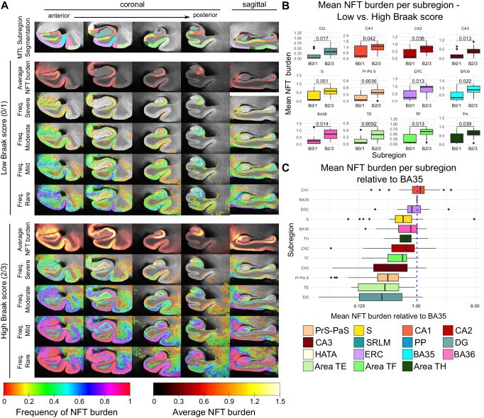
In a study in Nature Communications, Sadhana Ravikumar, Paul Yushkevich and colleagues combined ex vivo MRI with serial histological imaging of 25 human MTL specimens. They mapped quantitative NFT burden into a high-resolution ex vivo atlas whose subregions are defined by cytoarchitecture, creating a detailed 3D picture of where tangles accumulate.
Key findings:
- Average maps show a clear anterior-to-posterior gradient in NFT distribution.
- The highest tangle burden is found not only in the transentorhinal region, but also in the CA1 subfield of the hippocampus.
- The study identifies fine-grained MTL regions where neurodegeneration is likely to be linked specifically to tangles. Measurements there may be more sensitive early biomarkers of Alzheimer’s disease.
Other PICSL contributors include Amanda Denning, Sydney Lim, Ranjit Ittyerah, Laura Wisse, Sandhitsu Das and Long Xie.
Reference: Ravikumar S, Denning AE, Lim S, et al., Yushkevich PA. Postmortem imaging reveals patterns of medial temporal lobe vulnerability to tau pathology in Alzheimer’s disease. Nature Communications. 2024. doi:10.1038/s41467-024-49205-0