
An ex vivo atlas of the earliest brain regions affected by Alzheimer's disease

The earliest neuropathological changes of Alzheimer’s disease (AD) appear in the medial temporal lobe (MTL). For MRI biomarkers to detect changes caused specifically by AD, rather than by aging or other diseases, patterns of structural change in the MTL need to be linked to the underlying pathology.
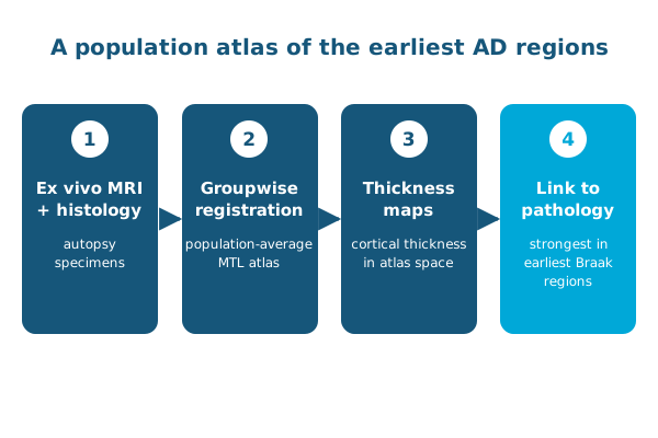
At the IEEE International Symposium on Biomedical Imaging (ISBI 2020), Sadhana Ravikumar, Paul Yushkevich and colleagues described an autopsy imaging study that combines ex vivo MRI with serial histopathology. To study many donors together, they developed a groupwise registration approach that builds a population-average atlas of the MTL, capturing both its complex structure and how it varies between people.
Using the atlas, they generated maps of cortical thickness and found a significant correlation between atrophy and AD pathology in the MTL subregions affected at the earliest stages of AD, as described by Braak and Braak. This work laid the foundation for the later 3D atlas of tau tangles in the MTL.
Other PICSL contributors include Laura Wisse, Ranjit Ittyerah, Sydney Lim, Long Xie and Sandhitsu Das.
Reference: Ravikumar S, Wisse L, Ittyerah R, et al., Yushkevich PA. Building an ex vivo atlas of the earliest brain regions affected by Alzheimer’s disease pathology. IEEE International Symposium on Biomedical Imaging (ISBI). 2020. doi:10.1109/ISBI45749.2020.9098427